< Back
Informasi Alat
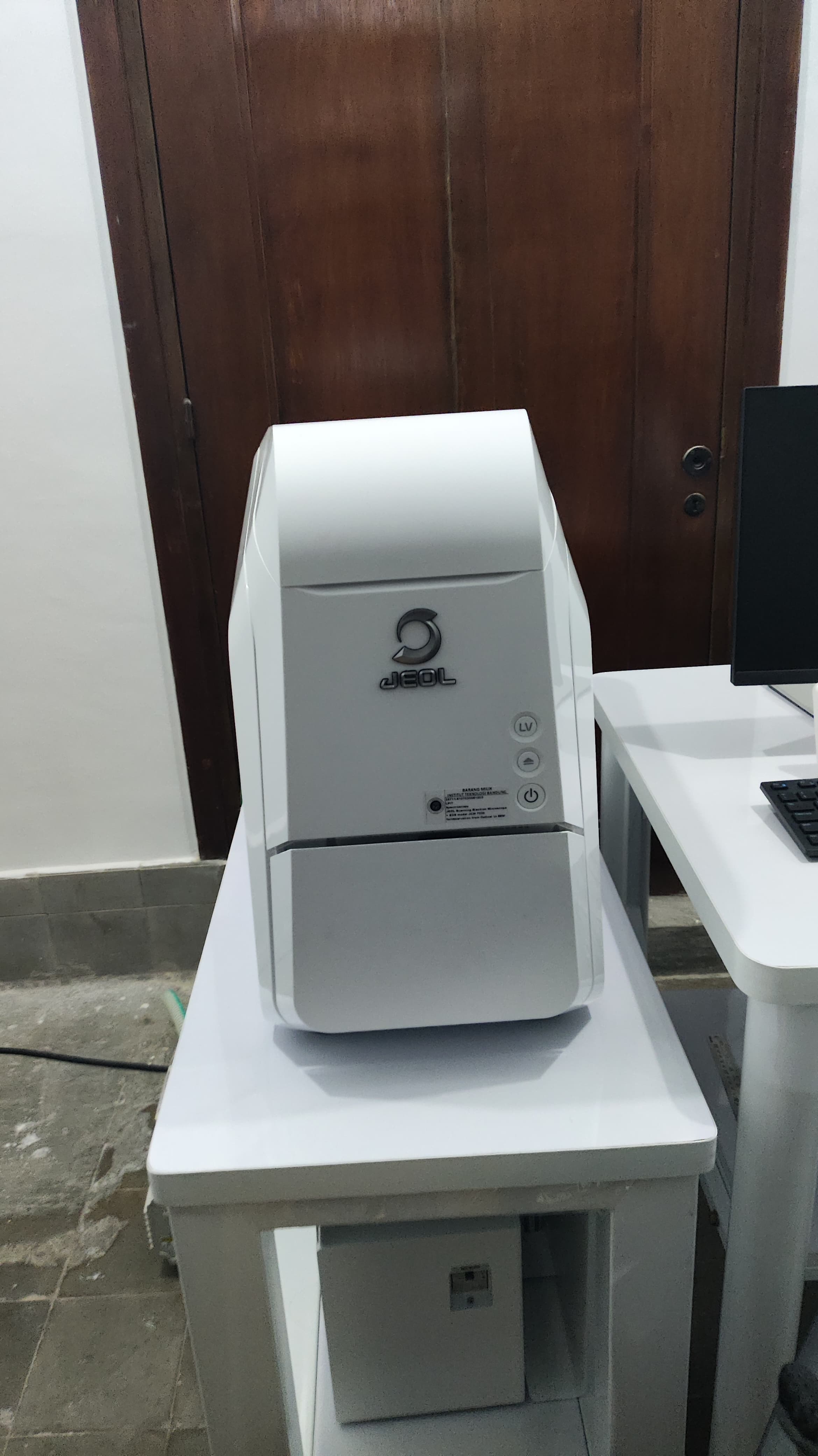
| Nama Alat | Scanning Electron Microscope |
|---|---|
| Deskripsi Alat | SEM-EDS is a powerful tool for material structure and elementer analysis. The related research topics are as follow: 1. Investigating new materials such as metal-organic frameworks (MOFs), zeolites, or amine-functionalized sorbents for their efficiency in capturing CO2. The SEM-EDS can be used to analyze the microstructure and elemental composition of these sorbents. 2. It can be also used for studying the surface and cross-sectional morphology of membrane materials used for CO2 separation. This includes assessing pore size, distribution, and the presence of active sites for CO2 adsorption. 3. Catalyst Development for CO2 Reduction: the surface properties and elemental composition of catalysts used in the chemical reduction of CO2 to valuable hydrocarbons. 4. Examining materials used in energy storage devices like batteries and supercapacitors, focusing on electrode materials, electrolytes, and interfaces. 5. Studying the surface characteristics and elemental composition of photocatalysts used in solar energy conversion, including water splitting and CO2 reduction to solar fuels. JEOLScanning Electron Microscope+EDS + COATER , model JCM7000 JCM-7000 NeoScope™ Benchtop SEM for observation from Optical to SEM image with direct Live Elemental Analysis SPECIFICATION : Electron Gun Electron source : - Precentered Tungsten filament, Automatic Gun Alignment (fully automatic alignment adjustment). - No tool required. - Changing the Filament is easy and fast, can be done by operator no need call engineer from agent. - Filament life time 100 hours Accelerating Voltage : 5, 10, 15 kV (3 steps switchover from the GUI). Magnification Image Magnification : ×10 to ×100,000 (magnification as defined by 128mm x 96mm ) Display Magnification : x24 to x202,168 (magnification as defined by 280 mm × 210 mm) Specimen stage Specimen movement : 2 axes (X, Y) Motor-driven Specimen movement range : X-direction = 40 mm; Y-direction = 40 mm Specimen holder : 32 mm (ɸ)× 44 mm (H) & 80 mm disc Maximum specimen size : 80 mm (ɸ)× 50 mm (H) Specimen exchange : Stage draw-out style mechanism Detector - High-vacuum mode : SEI Secondary Electron Image. BEI Backscattered Electron Image (Composition, topographic, stereoscopic and 3D images). - Low-vacuum mode :BEI Backscattered Electron Image (Composition, topographic, stereoscopic and 3D images). Pixel for Image function : 5,120 x 3,840 Measurement functions Two-point measurement (straight) : Measurement of the distance between the straight line segment connecting any two points and the length of a series of connected straight line segments. Angle measurement: Measurement of the angle between two line segments that extend from a center point. Line width measurement (parallel X, Y): Measures the distance between a reference line and a parallel line. File saving Image file format : Can be saved in BMP, TIFF, JPEG, or PNG. Computer Desktop PC i5 with 24” touch screen LCD monitor Evacuation System Control method : Fully automatic control TMP & RP SMART Fine Coater with Gold Target This coater for coating the surface of Non Conductive sample before running into SEM Stage Navigation System (SNS) use Color CCD Camera With SNS will get smoothing transition once image observation from optical to SEM Images and will be displayed directly in real time of EDS element analysis (Optical Image to SEM observation and direct EDS LIVE ANALYSIS information). SPECIFICATIONS FOR JEOL Dry EDS (Energy Dispersive Spectrometer) Detector (Liquid Nitrogen free) Sensor: Silicon drift detector (SDD), Detector element area: 30 mm2 , Window : Polymer thin film, Detectable element range : Be to U Energy resolution (Mn-Ka FWHM): 130 eV or less (ICR 3,000 cps or less), Cooling method: Peltier Cooling Usable accelerating voltage: 15kV or less Retraction mechanism: Not available (Non-retractable) Digital pulse processor (DPP6) The digital pulse processor digitally processes X-ray signals detected by the X-ray detector and outputs the X-ray energy signals scanned and synced with the JCM-7000. EDS Software capable : - Image Acquisition - Spectrum Acquisition - Qualitative Analysis - Quantitative Analysis - Line Measurement - X-Ray Mapping Consisting of : 1. JCM-7000 Benchtop Scanning Electron Microscope, Consisting of : BENCHTOP SEM Basic Unit of JCM-7000 Motorize Drive Stage with Axes X,Y Rotary Pump Attachment Control Unit Power supply Kits 5 Stage Navigation System (using color CCD Camera) EDS Kit Dry SD30 Detector Unit Basic Software SMART Coater TOOL Box 2. Accessories & Consumable Parts : Filament (V Type) = 6pcs Wehnelt = 1pc Carbon Tape = 1pc Vacuum Grease = 1pc Specimen Holder (32mmφ) = 1pc (including 2 specimen block) Specimen Holder (80mmφ)= 1pc (shaped like disk) Included Local Supply : Automatic Voltage Regulator UPS 1.2 kVA for PC Intel Processor i5, 8GB RAM, SSD 250GB, HDD 1TB, Windows 10, Touch Screen LCD Monitor 24” Ink Jet Color Printer A4 Table for SEM JCM-7000 |
| Preparasi Sample | |
| Lokasi | Ruang SEM, Lab Logam (Lab Metalurgi dan Teknik Material) FTMD ITB |
| PIC | Yorina Sarah F Lantang |
| Status | Active : Penggunaan Alat dapat dilakukan oleh semua users. |
| Waktu Penggunaan Alat | Per-jam |